
Image by the US National Institutes of Health, CC 3.0
A new dual-light microscope lets researchers observe micro- and nanoscale activity inside living cells without using dyes. The system, from the University of Tokyo, captures both detailed structures and tiny moving particles at once, providing a more complete view of cellular behaviour.
The technology is in the form of a microscope capable of detecting signals across an intensity range fourteen times broader than that of standard instruments. The gentle, dye-free approach allows cells to remain unharmed during long-term imaging, which could benefit testing and quality control in pharmaceutical and biotechnology settings.
Combining technologies
The microscope combines two advanced technologies: Quantitative phase microscopy and interferometric scattering microscopy
Quantitative phase microscopy (QPM) uses forward-scattered light to visualise structures at the microscale (in this study, over 100 nanometers), which makes it useful for capturing still images of complex cell features. In other words, the microscope quantifies the phase shift that occurs when light waves pass through a more optically dense object. However, QPM cannot detect very small particles.

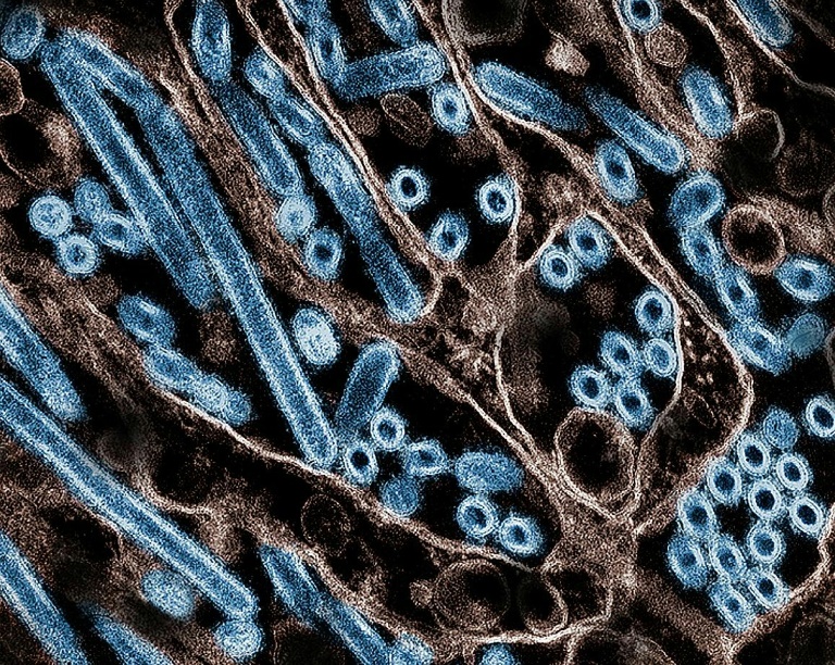
A colorized transmission electron micrograph of avian influenza A H5N1 virus particles (blue), grown in Madin-Darby Canine Kidney (MDCK) epithelial cells – Copyright National Institute of Allergy and Infectious Diseases/AFP/File HANDOUT
Interferometric scattering (iSCAT) microscopy works differently by capturing back-scattered light and can detect structures as tiny as single proteins. The key feature of iSCAT is the detection of elastic scattering from subwavelength particles, also known as Rayleigh scattering, in addition to reflected or transmission signals from supra-wavelength objects. While iSCAT enables researchers to “track” individual particles and observe rapid changes inside cells, it lacks the wider view offered by QPM.
The researchers examined whether collecting light from both directions simultaneously could bridge the gap and reveal activity across a broad range of sizes and motions in a single image.
The development is referred to as bidirectional quantitative scattering microscopy (BiQSM). This uses forward scattering and backward scattering approaches, using off-axis digital holography with bidirectional illumination and spatial-frequency multiplexing.
Exploration and realisation
To explore the idea and confirm that their microscope performed as expected, they observed how cells behaved during cell death. In one experiment, they captured an image that contained information from both forward- and backward-traveling light.
The device was tested by analysing changes during cell death. Through this, the team were able to estimate particle size and refractive index. The scientists hope to push the technique toward imaging particles as small as viruses.
The researchers had succeeded in identifying the movement of larger cell structures (micro) as well as much smaller particles (nano). By comparing the patterns in forward- and back-scattered light, they could estimate each particle’s size and its refractive index, which describes how strongly light bends or scatters when it passes through a material.
The development is described in the journal Nature Biotechnology, with the paper titled “Bidirectional quantitative scattering microscopy.”


